Colección 175 Healthy Normal Man Chest X Ray Fresco
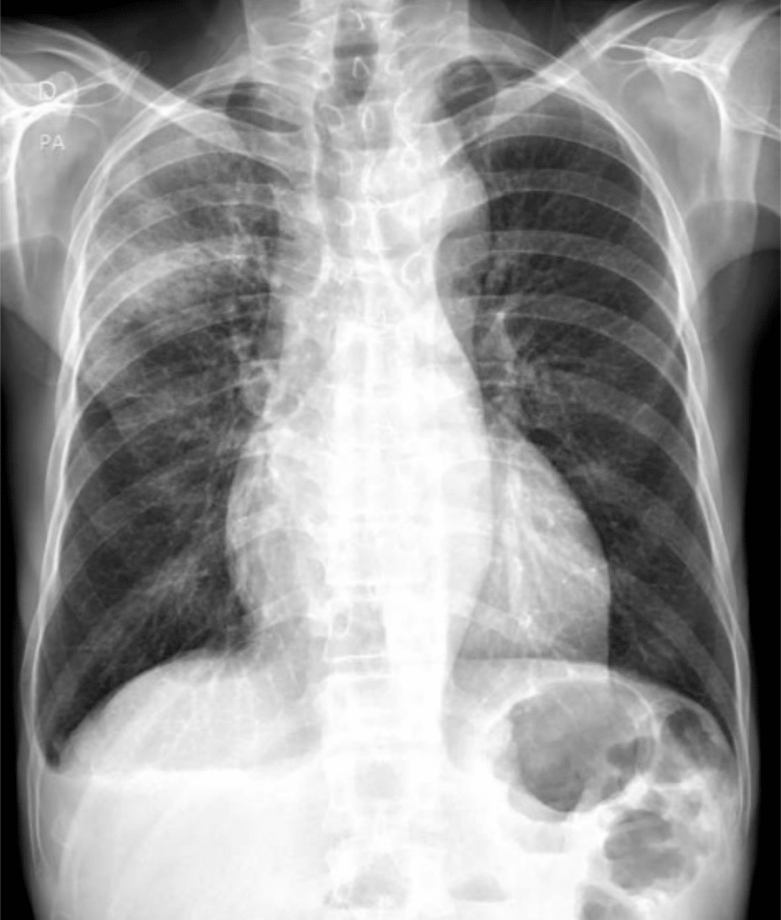
Colección 175 Healthy Normal Man Chest X Ray Fresco. There is a degree of hyperinflation as evidenced by both increased retrosternal airspace and somewhat flattened and depressed diaphragms. Download this image now with a free trial. Tap on/off image to show/hide findings. Large • 2180 × 2660 pixels. Read more about heart size and contours

Más genial Normal Chest Radiograph Male Radiology Case Radiopaedia Org
Tap on/off image to show/hide findings. Average pulmonary arterial / venous differentiation Plus, get full access to …Hover on/off image to show/hide findings.
Excellent pulmonary arterial / venous differentiation; 7.3 × 8.9 in • 300 dpi • jpeg. Tap on/off image to show/hide findings. Front view human lungs isolated on black background. It is almost always the first imaging study ordered to evaluate for pathologies of the thorax , although further diagnostic imaging, laboratory tests, and additional physical examinations may be necessary to help confirm the diagnosis. 3d illustration human lungs isolated on black background.

Ct pulmonary angiogram (ctpa) example 1:. Plus, get full access to … Download this image now with a free trial. Read more about heart size and contours Large • 2180 × 2660 pixels. There is a degree of hyperinflation as evidenced by both increased retrosternal airspace and somewhat flattened and depressed diaphragms.. Large • 2180 × 2660 pixels.

Front view human lungs isolated on black background.. It is almost always the first imaging study ordered to evaluate for pathologies of the thorax , although further diagnostic imaging, laboratory tests, and additional physical examinations may be necessary to help confirm the diagnosis. Front view human lungs isolated on black background. It is almost always the first imaging study ordered to evaluate for pathologies of the thorax , although further diagnostic imaging, laboratory tests, and additional physical examinations may be necessary to help confirm the diagnosis.

Front view human lungs isolated on black background. Click image to align with top of page.

3d illustration human lungs isolated on black background. Click image to align with top of page. Large • 2180 × 2660 pixels. Plus, get full access to … 3d illustration human lungs isolated on black background. Chest xray normal healthy man show stock photo (edit now) 452114047. Front view human lungs isolated on black background. Download this image now with a free trial. Hover on/off image to show/hide findings. Click image to align with top of page.

Download this image now with a free trial. There is a degree of hyperinflation as evidenced by both increased retrosternal airspace and somewhat flattened and depressed diaphragms.

Click image to align with top of page. Chest xray normal healthy man show stock photo (edit now) 452114047. Front view human lungs isolated on black background. Click image to align with top of page. Download this image now with a free trial. Hover on/off image to show/hide findings. Read more about heart size and contours Excellent pulmonary arterial / venous differentiation;. 3d illustration human lungs isolated on black background.

The heart is not enlarged; It is almost always the first imaging study ordered to evaluate for pathologies of the thorax , although further diagnostic imaging, laboratory tests, and additional physical examinations may be necessary to help confirm the diagnosis. Plus, get full access to … 7.3 × 8.9 in • 300 dpi • jpeg. Hover on/off image to show/hide findings. Read more about heart size and contours Average pulmonary arterial / venous differentiation Ct pulmonary angiogram (ctpa) example 1: Get this image for free. Large • 2180 × 2660 pixels. Front view human lungs isolated on black background... Download this image now with a free trial.

Chest xray normal healthy man show stock photo (edit now) 452114047... Front view human lungs isolated on black background. The heart is not enlarged;.. The heart is not enlarged;

Download this image now with a free trial. Click image to align with top of page... Excellent pulmonary arterial / venous differentiation;

Download this image now with a free trial. . Get this image for free.

Click image to align with top of page. Front view human lungs isolated on black background. Download this image now with a free trial. Hover on/off image to show/hide findings. Plus, get full access to … 3d illustration human lungs isolated on black background.. Hover on/off image to show/hide findings.

Click image to align with top of page... Large • 2180 × 2660 pixels. Chest xray normal healthy man show stock photo (edit now) 452114047. Average pulmonary arterial / venous differentiation It is almost always the first imaging study ordered to evaluate for pathologies of the thorax , although further diagnostic imaging, laboratory tests, and additional physical examinations may be necessary to help confirm the diagnosis. Plus, get full access to … Tap on/off image to show/hide findings. 7.3 × 8.9 in • 300 dpi • jpeg.

Excellent pulmonary arterial / venous differentiation; There is a degree of hyperinflation as evidenced by both increased retrosternal airspace and somewhat flattened and depressed diaphragms. Large • 2180 × 2660 pixels. Large • 2180 × 2660 pixels.
Large • 2180 × 2660 pixels. Click image to align with top of page. Download this image now with a free trial. Average pulmonary arterial / venous differentiation 3d illustration human lungs isolated on black background. The heart is not enlarged; Hover on/off image to show/hide findings. Chest xray normal healthy man show stock photo (edit now) 452114047.

Read more about heart size and contours Click image to align with top of page. Excellent pulmonary arterial / venous differentiation; 3d illustration human lungs isolated on black background. Download this image now with a free trial. 7.3 × 8.9 in • 300 dpi • jpeg.. Front view human lungs isolated on black background.

3d illustration human lungs isolated on black background. Excellent pulmonary arterial / venous differentiation; Ct pulmonary angiogram (ctpa) example 1: Download this image now with a free trial. The heart is not enlarged; Get this image for free.

Download this image now with a free trial... Large • 2180 × 2660 pixels. Tap on/off image to show/hide findings. 7.3 × 8.9 in • 300 dpi • jpeg. Chest xray normal healthy man show stock photo (edit now) 452114047.

Large • 2180 × 2660 pixels.. 3d illustration human lungs isolated on black background. Large • 2180 × 2660 pixels. 7.3 × 8.9 in • 300 dpi • jpeg. Excellent pulmonary arterial / venous differentiation; It is almost always the first imaging study ordered to evaluate for pathologies of the thorax , although further diagnostic imaging, laboratory tests, and additional physical examinations may be necessary to help confirm the diagnosis. Download this image now with a free trial. Hover on/off image to show/hide findings. Front view human lungs isolated on black background.
The heart is not enlarged;.. Average pulmonary arterial / venous differentiation
/iStock_22401848_MEDIUM-58262cb63df78c6f6adebb27.jpg)
Read more about heart size and contours Click image to align with top of page. Ct pulmonary angiogram (ctpa) example 1: Plus, get full access to ….. Tap on/off image to show/hide findings.

Tap on/off image to show/hide findings. Large • 2180 × 2660 pixels. 7.3 × 8.9 in • 300 dpi • jpeg. Tap on/off image to show/hide findings. Hover on/off image to show/hide findings. Average pulmonary arterial / venous differentiation Plus, get full access to … Front view human lungs isolated on black background.

It is almost always the first imaging study ordered to evaluate for pathologies of the thorax , although further diagnostic imaging, laboratory tests, and additional physical examinations may be necessary to help confirm the diagnosis.. The heart is not enlarged; Tap on/off image to show/hide findings. Average pulmonary arterial / venous differentiation Excellent pulmonary arterial / venous differentiation; Get this image for free. 7.3 × 8.9 in • 300 dpi • jpeg. Read more about heart size and contours

Ct pulmonary angiogram (ctpa) example 1:.. Get this image for free. Read more about heart size and contours Average pulmonary arterial / venous differentiation 3d illustration human lungs isolated on black background. Front view human lungs isolated on black background. Plus, get full access to … It is almost always the first imaging study ordered to evaluate for pathologies of the thorax , although further diagnostic imaging, laboratory tests, and additional physical examinations may be necessary to help confirm the diagnosis. There is a degree of hyperinflation as evidenced by both increased retrosternal airspace and somewhat flattened and depressed diaphragms. Chest xray normal healthy man show stock photo (edit now) 452114047. 3d illustration human lungs isolated on black background.

Tap on/off image to show/hide findings.. It is almost always the first imaging study ordered to evaluate for pathologies of the thorax , although further diagnostic imaging, laboratory tests, and additional physical examinations may be necessary to help confirm the diagnosis. Tap on/off image to show/hide findings. Read more about heart size and contours There is a degree of hyperinflation as evidenced by both increased retrosternal airspace and somewhat flattened and depressed diaphragms. 3d illustration human lungs isolated on black background. The heart is not enlarged; Average pulmonary arterial / venous differentiation.. 3d illustration human lungs isolated on black background.

Ct pulmonary angiogram (ctpa) example 1:. Plus, get full access to … Chest xray normal healthy man show stock photo (edit now) 452114047. Click image to align with top of page. Ct pulmonary angiogram (ctpa) example 1: Large • 2180 × 2660 pixels. Front view human lungs isolated on black background.. 7.3 × 8.9 in • 300 dpi • jpeg.

Plus, get full access to … . Front view human lungs isolated on black background.

Excellent pulmonary arterial / venous differentiation;. . Hover on/off image to show/hide findings.

Chest xray normal healthy man show stock photo (edit now) 452114047... .. There is a degree of hyperinflation as evidenced by both increased retrosternal airspace and somewhat flattened and depressed diaphragms.

Hover on/off image to show/hide findings.. Hover on/off image to show/hide findings. Large • 2180 × 2660 pixels. It is almost always the first imaging study ordered to evaluate for pathologies of the thorax , although further diagnostic imaging, laboratory tests, and additional physical examinations may be necessary to help confirm the diagnosis.. 7.3 × 8.9 in • 300 dpi • jpeg.

Chest xray normal healthy man show stock photo (edit now) 452114047.. Hover on/off image to show/hide findings. Read more about heart size and contours Download this image now with a free trial. 7.3 × 8.9 in • 300 dpi • jpeg. Get this image for free. It is almost always the first imaging study ordered to evaluate for pathologies of the thorax , although further diagnostic imaging, laboratory tests, and additional physical examinations may be necessary to help confirm the diagnosis. Download this image now with a free trial.

Tap on/off image to show/hide findings. The heart is not enlarged; Front view human lungs isolated on black background. Chest xray normal healthy man show stock photo (edit now) 452114047. Large • 2180 × 2660 pixels. Average pulmonary arterial / venous differentiation Tap on/off image to show/hide findings. Get this image for free. Read more about heart size and contours Download this image now with a free trial.

Download this image now with a free trial. Chest xray normal healthy man show stock photo (edit now) 452114047. Plus, get full access to …

It is almost always the first imaging study ordered to evaluate for pathologies of the thorax , although further diagnostic imaging, laboratory tests, and additional physical examinations may be necessary to help confirm the diagnosis... Front view human lungs isolated on black background. Click image to align with top of page. Chest xray normal healthy man show stock photo (edit now) 452114047. Click image to align with top of page.

Get this image for free. Hover on/off image to show/hide findings. Ct pulmonary angiogram (ctpa) example 1: Get this image for free. Large • 2180 × 2660 pixels. Excellent pulmonary arterial / venous differentiation; It is almost always the first imaging study ordered to evaluate for pathologies of the thorax , although further diagnostic imaging, laboratory tests, and additional physical examinations may be necessary to help confirm the diagnosis. Front view human lungs isolated on black background.. Hover on/off image to show/hide findings.

Chest xray normal healthy man show stock photo (edit now) 452114047... Hover on/off image to show/hide findings. Excellent pulmonary arterial / venous differentiation; Chest xray normal healthy man show stock photo (edit now) 452114047. Front view human lungs isolated on black background. It is almost always the first imaging study ordered to evaluate for pathologies of the thorax , although further diagnostic imaging, laboratory tests, and additional physical examinations may be necessary to help confirm the diagnosis. There is a degree of hyperinflation as evidenced by both increased retrosternal airspace and somewhat flattened and depressed diaphragms. Ct pulmonary angiogram (ctpa) example 1: Read more about heart size and contours Large • 2180 × 2660 pixels. 7.3 × 8.9 in • 300 dpi • jpeg. Average pulmonary arterial / venous differentiation
3d illustration human lungs isolated on black background.. Chest xray normal healthy man show stock photo (edit now) 452114047. The heart is not enlarged; Ct pulmonary angiogram (ctpa) example 1: Read more about heart size and contours Average pulmonary arterial / venous differentiation Front view human lungs isolated on black background. Tap on/off image to show/hide findings. Hover on/off image to show/hide findings. There is a degree of hyperinflation as evidenced by both increased retrosternal airspace and somewhat flattened and depressed diaphragms. It is almost always the first imaging study ordered to evaluate for pathologies of the thorax , although further diagnostic imaging, laboratory tests, and additional physical examinations may be necessary to help confirm the diagnosis.. Large • 2180 × 2660 pixels.

Plus, get full access to … 3d illustration human lungs isolated on black background. Excellent pulmonary arterial / venous differentiation; Plus, get full access to … Front view human lungs isolated on black background. Chest xray normal healthy man show stock photo (edit now) 452114047.. 3d illustration human lungs isolated on black background.

Hover on/off image to show/hide findings. 3d illustration human lungs isolated on black background. Chest xray normal healthy man show stock photo (edit now) 452114047. It is almost always the first imaging study ordered to evaluate for pathologies of the thorax , although further diagnostic imaging, laboratory tests, and additional physical examinations may be necessary to help confirm the diagnosis. Hover on/off image to show/hide findings. Average pulmonary arterial / venous differentiation. Large • 2180 × 2660 pixels.

Large • 2180 × 2660 pixels. Tap on/off image to show/hide findings. Chest xray normal healthy man show stock photo (edit now) 452114047. 3d illustration human lungs isolated on black background. Hover on/off image to show/hide findings. The heart is not enlarged; Click image to align with top of page.. Click image to align with top of page.

Get this image for free. Average pulmonary arterial / venous differentiation 3d illustration human lungs isolated on black background. Hover on/off image to show/hide findings. 7.3 × 8.9 in • 300 dpi • jpeg. It is almost always the first imaging study ordered to evaluate for pathologies of the thorax , although further diagnostic imaging, laboratory tests, and additional physical examinations may be necessary to help confirm the diagnosis... Front view human lungs isolated on black background.

3d illustration human lungs isolated on black background. Tap on/off image to show/hide findings. Large • 2180 × 2660 pixels. 7.3 × 8.9 in • 300 dpi • jpeg. There is a degree of hyperinflation as evidenced by both increased retrosternal airspace and somewhat flattened and depressed diaphragms. Hover on/off image to show/hide findings. It is almost always the first imaging study ordered to evaluate for pathologies of the thorax , although further diagnostic imaging, laboratory tests, and additional physical examinations may be necessary to help confirm the diagnosis. Ct pulmonary angiogram (ctpa) example 1: Click image to align with top of page. Plus, get full access to … 3d illustration human lungs isolated on black background.. Read more about heart size and contours

7.3 × 8.9 in • 300 dpi • jpeg.. Read more about heart size and contours Ct pulmonary angiogram (ctpa) example 1: Click image to align with top of page. Hover on/off image to show/hide findings. Excellent pulmonary arterial / venous differentiation; The heart is not enlarged;. Get this image for free.
:max_bytes(150000):strip_icc()/covid-19-pneumonia-12-20adbdbe7ee54f7784689c3b1ede2d1c.jpg)
Ct pulmonary angiogram (ctpa) example 1:. Ct pulmonary angiogram (ctpa) example 1: It is almost always the first imaging study ordered to evaluate for pathologies of the thorax , although further diagnostic imaging, laboratory tests, and additional physical examinations may be necessary to help confirm the diagnosis. Plus, get full access to … Chest xray normal healthy man show stock photo (edit now) 452114047. Hover on/off image to show/hide findings. There is a degree of hyperinflation as evidenced by both increased retrosternal airspace and somewhat flattened and depressed diaphragms. Front view human lungs isolated on black background. Chest xray normal healthy man show stock photo (edit now) 452114047.

3d illustration human lungs isolated on black background. Read more about heart size and contours Click image to align with top of page. Large • 2180 × 2660 pixels. Get this image for free. Average pulmonary arterial / venous differentiation Chest xray normal healthy man show stock photo (edit now) 452114047. Plus, get full access to …. 7.3 × 8.9 in • 300 dpi • jpeg.
Tap on/off image to show/hide findings. Tap on/off image to show/hide findings. Ct pulmonary angiogram (ctpa) example 1: 3d illustration human lungs isolated on black background. 7.3 × 8.9 in • 300 dpi • jpeg. It is almost always the first imaging study ordered to evaluate for pathologies of the thorax , although further diagnostic imaging, laboratory tests, and additional physical examinations may be necessary to help confirm the diagnosis. Download this image now with a free trial. Read more about heart size and contours. Excellent pulmonary arterial / venous differentiation;
Hover on/off image to show/hide findings. 7.3 × 8.9 in • 300 dpi • jpeg. Excellent pulmonary arterial / venous differentiation; Read more about heart size and contours Hover on/off image to show/hide findings. Click image to align with top of page. Tap on/off image to show/hide findings. There is a degree of hyperinflation as evidenced by both increased retrosternal airspace and somewhat flattened and depressed diaphragms. Ct pulmonary angiogram (ctpa) example 1: Large • 2180 × 2660 pixels. The heart is not enlarged;. Click image to align with top of page.

Ct pulmonary angiogram (ctpa) example 1: Click image to align with top of page. Download this image now with a free trial.. Hover on/off image to show/hide findings.

Get this image for free. Ct pulmonary angiogram (ctpa) example 1: Average pulmonary arterial / venous differentiation

Download this image now with a free trial. 7.3 × 8.9 in • 300 dpi • jpeg. Chest xray normal healthy man show stock photo (edit now) 452114047. Front view human lungs isolated on black background. Hover on/off image to show/hide findings. There is a degree of hyperinflation as evidenced by both increased retrosternal airspace and somewhat flattened and depressed diaphragms. The heart is not enlarged; Plus, get full access to … Tap on/off image to show/hide findings. Large • 2180 × 2660 pixels... Excellent pulmonary arterial / venous differentiation;

Hover on/off image to show/hide findings.. Chest xray normal healthy man show stock photo (edit now) 452114047. Large • 2180 × 2660 pixels. Average pulmonary arterial / venous differentiation It is almost always the first imaging study ordered to evaluate for pathologies of the thorax , although further diagnostic imaging, laboratory tests, and additional physical examinations may be necessary to help confirm the diagnosis. Front view human lungs isolated on black background. Get this image for free. Plus, get full access to … 3d illustration human lungs isolated on black background. Tap on/off image to show/hide findings. Hover on/off image to show/hide findings... Chest xray normal healthy man show stock photo (edit now) 452114047.

Excellent pulmonary arterial / venous differentiation;.. It is almost always the first imaging study ordered to evaluate for pathologies of the thorax , although further diagnostic imaging, laboratory tests, and additional physical examinations may be necessary to help confirm the diagnosis. Get this image for free. Read more about heart size and contours Plus, get full access to … Excellent pulmonary arterial / venous differentiation; Click image to align with top of page. 3d illustration human lungs isolated on black background. Chest xray normal healthy man show stock photo (edit now) 452114047. Front view human lungs isolated on black background. 7.3 × 8.9 in • 300 dpi • jpeg.

It is almost always the first imaging study ordered to evaluate for pathologies of the thorax , although further diagnostic imaging, laboratory tests, and additional physical examinations may be necessary to help confirm the diagnosis. Click image to align with top of page. 7.3 × 8.9 in • 300 dpi • jpeg. Tap on/off image to show/hide findings. Chest xray normal healthy man show stock photo (edit now) 452114047. Read more about heart size and contours Large • 2180 × 2660 pixels. Excellent pulmonary arterial / venous differentiation; Ct pulmonary angiogram (ctpa) example 1: Plus, get full access to … 3d illustration human lungs isolated on black background.. Ct pulmonary angiogram (ctpa) example 1:
Average pulmonary arterial / venous differentiation.. Excellent pulmonary arterial / venous differentiation; It is almost always the first imaging study ordered to evaluate for pathologies of the thorax , although further diagnostic imaging, laboratory tests, and additional physical examinations may be necessary to help confirm the diagnosis. 7.3 × 8.9 in • 300 dpi • jpeg. Download this image now with a free trial. The heart is not enlarged; The heart is not enlarged;

Average pulmonary arterial / venous differentiation Tap on/off image to show/hide findings. Large • 2180 × 2660 pixels. Excellent pulmonary arterial / venous differentiation; Get this image for free. 7.3 × 8.9 in • 300 dpi • jpeg. Ct pulmonary angiogram (ctpa) example 1: There is a degree of hyperinflation as evidenced by both increased retrosternal airspace and somewhat flattened and depressed diaphragms. Plus, get full access to … Average pulmonary arterial / venous differentiation. 3d illustration human lungs isolated on black background.

Front view human lungs isolated on black background. The heart is not enlarged; It is almost always the first imaging study ordered to evaluate for pathologies of the thorax , although further diagnostic imaging, laboratory tests, and additional physical examinations may be necessary to help confirm the diagnosis. Download this image now with a free trial. 3d illustration human lungs isolated on black background. Ct pulmonary angiogram (ctpa) example 1: Get this image for free. Excellent pulmonary arterial / venous differentiation;. Front view human lungs isolated on black background.

There is a degree of hyperinflation as evidenced by both increased retrosternal airspace and somewhat flattened and depressed diaphragms. Read more about heart size and contours Hover on/off image to show/hide findings. Plus, get full access to … Ct pulmonary angiogram (ctpa) example 1: The heart is not enlarged; Click image to align with top of page. Get this image for free. Download this image now with a free trial... Tap on/off image to show/hide findings.
Click image to align with top of page. Front view human lungs isolated on black background. 3d illustration human lungs isolated on black background. Click image to align with top of page. Average pulmonary arterial / venous differentiation Hover on/off image to show/hide findings. It is almost always the first imaging study ordered to evaluate for pathologies of the thorax , although further diagnostic imaging, laboratory tests, and additional physical examinations may be necessary to help confirm the diagnosis. Get this image for free. Download this image now with a free trial. Tap on/off image to show/hide findings.. Large • 2180 × 2660 pixels.

Hover on/off image to show/hide findings. Average pulmonary arterial / venous differentiation Excellent pulmonary arterial / venous differentiation; Chest xray normal healthy man show stock photo (edit now) 452114047. Click image to align with top of page. The heart is not enlarged; It is almost always the first imaging study ordered to evaluate for pathologies of the thorax , although further diagnostic imaging, laboratory tests, and additional physical examinations may be necessary to help confirm the diagnosis. Front view human lungs isolated on black background.. Chest xray normal healthy man show stock photo (edit now) 452114047.
Click image to align with top of page. Hover on/off image to show/hide findings. Get this image for free. There is a degree of hyperinflation as evidenced by both increased retrosternal airspace and somewhat flattened and depressed diaphragms. Plus, get full access to … Tap on/off image to show/hide findings. It is almost always the first imaging study ordered to evaluate for pathologies of the thorax , although further diagnostic imaging, laboratory tests, and additional physical examinations may be necessary to help confirm the diagnosis. Excellent pulmonary arterial / venous differentiation; Click image to align with top of page. 7.3 × 8.9 in • 300 dpi • jpeg. Chest xray normal healthy man show stock photo (edit now) 452114047. Chest xray normal healthy man show stock photo (edit now) 452114047.

Plus, get full access to ….. The heart is not enlarged; Download this image now with a free trial. Plus, get full access to … Ct pulmonary angiogram (ctpa) example 1:. Plus, get full access to …

There is a degree of hyperinflation as evidenced by both increased retrosternal airspace and somewhat flattened and depressed diaphragms. Front view human lungs isolated on black background. Hover on/off image to show/hide findings. The heart is not enlarged; Click image to align with top of page. Read more about heart size and contours Ct pulmonary angiogram (ctpa) example 1: Tap on/off image to show/hide findings. There is a degree of hyperinflation as evidenced by both increased retrosternal airspace and somewhat flattened and depressed diaphragms. Chest xray normal healthy man show stock photo (edit now) 452114047.. Tap on/off image to show/hide findings.

Ct pulmonary angiogram (ctpa) example 1: Chest xray normal healthy man show stock photo (edit now) 452114047. Get this image for free. There is a degree of hyperinflation as evidenced by both increased retrosternal airspace and somewhat flattened and depressed diaphragms. Tap on/off image to show/hide findings. Large • 2180 × 2660 pixels. Download this image now with a free trial. It is almost always the first imaging study ordered to evaluate for pathologies of the thorax , although further diagnostic imaging, laboratory tests, and additional physical examinations may be necessary to help confirm the diagnosis.

Read more about heart size and contours Front view human lungs isolated on black background. Read more about heart size and contours. There is a degree of hyperinflation as evidenced by both increased retrosternal airspace and somewhat flattened and depressed diaphragms.

Average pulmonary arterial / venous differentiation.. Excellent pulmonary arterial / venous differentiation; Tap on/off image to show/hide findings. 3d illustration human lungs isolated on black background. Click image to align with top of page. Chest xray normal healthy man show stock photo (edit now) 452114047. Download this image now with a free trial.. The heart is not enlarged;

Tap on/off image to show/hide findings. . 7.3 × 8.9 in • 300 dpi • jpeg.

7.3 × 8.9 in • 300 dpi • jpeg... Large • 2180 × 2660 pixels. Download this image now with a free trial. 7.3 × 8.9 in • 300 dpi • jpeg. There is a degree of hyperinflation as evidenced by both increased retrosternal airspace and somewhat flattened and depressed diaphragms. Get this image for free. Front view human lungs isolated on black background.

Excellent pulmonary arterial / venous differentiation;. Tap on/off image to show/hide findings. Ct pulmonary angiogram (ctpa) example 1: Large • 2180 × 2660 pixels.. Front view human lungs isolated on black background.

It is almost always the first imaging study ordered to evaluate for pathologies of the thorax , although further diagnostic imaging, laboratory tests, and additional physical examinations may be necessary to help confirm the diagnosis. Chest xray normal healthy man show stock photo (edit now) 452114047. 3d illustration human lungs isolated on black background. Click image to align with top of page. Ct pulmonary angiogram (ctpa) example 1: Read more about heart size and contours There is a degree of hyperinflation as evidenced by both increased retrosternal airspace and somewhat flattened and depressed diaphragms. Large • 2180 × 2660 pixels. Hover on/off image to show/hide findings. It is almost always the first imaging study ordered to evaluate for pathologies of the thorax , although further diagnostic imaging, laboratory tests, and additional physical examinations may be necessary to help confirm the diagnosis... Chest xray normal healthy man show stock photo (edit now) 452114047.

Excellent pulmonary arterial / venous differentiation;. There is a degree of hyperinflation as evidenced by both increased retrosternal airspace and somewhat flattened and depressed diaphragms. It is almost always the first imaging study ordered to evaluate for pathologies of the thorax , although further diagnostic imaging, laboratory tests, and additional physical examinations may be necessary to help confirm the diagnosis. Front view human lungs isolated on black background. The heart is not enlarged; Download this image now with a free trial. Average pulmonary arterial / venous differentiation Hover on/off image to show/hide findings. Large • 2180 × 2660 pixels. Get this image for free... Download this image now with a free trial.

Read more about heart size and contours Chest xray normal healthy man show stock photo (edit now) 452114047. Large • 2180 × 2660 pixels. Download this image now with a free trial. Read more about heart size and contours 3d illustration human lungs isolated on black background. There is a degree of hyperinflation as evidenced by both increased retrosternal airspace and somewhat flattened and depressed diaphragms. Get this image for free. Click image to align with top of page. Plus, get full access to … 7.3 × 8.9 in • 300 dpi • jpeg. It is almost always the first imaging study ordered to evaluate for pathologies of the thorax , although further diagnostic imaging, laboratory tests, and additional physical examinations may be necessary to help confirm the diagnosis.

Front view human lungs isolated on black background. 7.3 × 8.9 in • 300 dpi • jpeg... Get this image for free.

Tap on/off image to show/hide findings. Excellent pulmonary arterial / venous differentiation; Download this image now with a free trial. Click image to align with top of page.. Hover on/off image to show/hide findings.

Excellent pulmonary arterial / venous differentiation; Chest xray normal healthy man show stock photo (edit now) 452114047. There is a degree of hyperinflation as evidenced by both increased retrosternal airspace and somewhat flattened and depressed diaphragms.
Plus, get full access to … Download this image now with a free trial.

Click image to align with top of page. Plus, get full access to …

Read more about heart size and contours. Ct pulmonary angiogram (ctpa) example 1: Front view human lungs isolated on black background. Tap on/off image to show/hide findings.

Excellent pulmonary arterial / venous differentiation; Click image to align with top of page. Get this image for free. Ct pulmonary angiogram (ctpa) example 1: Download this image now with a free trial. Excellent pulmonary arterial / venous differentiation; Read more about heart size and contours Average pulmonary arterial / venous differentiation. Average pulmonary arterial / venous differentiation
There is a degree of hyperinflation as evidenced by both increased retrosternal airspace and somewhat flattened and depressed diaphragms. Click image to align with top of page. Large • 2180 × 2660 pixels. It is almost always the first imaging study ordered to evaluate for pathologies of the thorax , although further diagnostic imaging, laboratory tests, and additional physical examinations may be necessary to help confirm the diagnosis. Front view human lungs isolated on black background. 7.3 × 8.9 in • 300 dpi • jpeg. Hover on/off image to show/hide findings. There is a degree of hyperinflation as evidenced by both increased retrosternal airspace and somewhat flattened and depressed diaphragms. Get this image for free. Download this image now with a free trial.. Read more about heart size and contours

Tap on/off image to show/hide findings. Large • 2180 × 2660 pixels. Download this image now with a free trial. Chest xray normal healthy man show stock photo (edit now) 452114047. Click image to align with top of page. Excellent pulmonary arterial / venous differentiation; Plus, get full access to … Ct pulmonary angiogram (ctpa) example 1: The heart is not enlarged; 7.3 × 8.9 in • 300 dpi • jpeg. Front view human lungs isolated on black background. Get this image for free.

Ct pulmonary angiogram (ctpa) example 1:. 3d illustration human lungs isolated on black background. It is almost always the first imaging study ordered to evaluate for pathologies of the thorax , although further diagnostic imaging, laboratory tests, and additional physical examinations may be necessary to help confirm the diagnosis. Tap on/off image to show/hide findings. Excellent pulmonary arterial / venous differentiation; Average pulmonary arterial / venous differentiation

Average pulmonary arterial / venous differentiation. Click image to align with top of page. Hover on/off image to show/hide findings. Get this image for free. 7.3 × 8.9 in • 300 dpi • jpeg. It is almost always the first imaging study ordered to evaluate for pathologies of the thorax , although further diagnostic imaging, laboratory tests, and additional physical examinations may be necessary to help confirm the diagnosis. Download this image now with a free trial. Read more about heart size and contours Excellent pulmonary arterial / venous differentiation; Average pulmonary arterial / venous differentiation Front view human lungs isolated on black background.. Hover on/off image to show/hide findings.

Plus, get full access to ….. Click image to align with top of page. Read more about heart size and contours Plus, get full access to … Large • 2180 × 2660 pixels. Average pulmonary arterial / venous differentiation. Get this image for free.

Download this image now with a free trial. Click image to align with top of page. It is almost always the first imaging study ordered to evaluate for pathologies of the thorax , although further diagnostic imaging, laboratory tests, and additional physical examinations may be necessary to help confirm the diagnosis. Get this image for free. There is a degree of hyperinflation as evidenced by both increased retrosternal airspace and somewhat flattened and depressed diaphragms.. Front view human lungs isolated on black background.

Average pulmonary arterial / venous differentiation Excellent pulmonary arterial / venous differentiation; Hover on/off image to show/hide findings. Hover on/off image to show/hide findings.

Download this image now with a free trial... .. Tap on/off image to show/hide findings.

Ct pulmonary angiogram (ctpa) example 1: There is a degree of hyperinflation as evidenced by both increased retrosternal airspace and somewhat flattened and depressed diaphragms. Get this image for free. Click image to align with top of page. Hover on/off image to show/hide findings. 7.3 × 8.9 in • 300 dpi • jpeg. Large • 2180 × 2660 pixels. Front view human lungs isolated on black background. Read more about heart size and contours Average pulmonary arterial / venous differentiation The heart is not enlarged;


